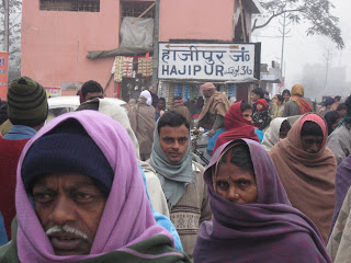
Leishmaniasis is a protozoal infection that is transmitted by phlebotomine sandflies. The disease is usually classified into two main groups: cutaneous leishmaniaisis (CL), which causes a skin rash or ulcer, and visceral leishmaniasis (VL), a disseminated form of the disease which causes a systemic febrile illness. VL is sometimes called Kala-Azar, which means “black sickness” in Hindi. Most CL is found in Central/South America and the Middle East, whereas most VL is in South Asia and Africa. More than 90% of the world’s kala-azar cases are in India, Bangladesh, Nepal, Brazil, (about 60% of cases in India, Bangladesh, or Nepal). More than 90% of Indian cases occur in Bihar State, the Kala-Azar capital of the world!
The CDC cartoon above diagrams the leishmaniasis lifecycle. When an infected sandfly bites a human, it injects the promastigote form of the protozoa. The promastigotes enter macrophages in the blood, then change into the round amastigote form. Infected macrophages travel to the spleen, lymph nodes, bone marrow, and other organs. The amastigote form multiplies inside the macrophages. Infected macrophages eventually burst, releasing amastigotes into the tissue. When a non-infected sandfly feeds on an infected patient, it ingests macrophages filled with amastigotes. The amastigotes turn into promastigotes in the gut of the sandfly. Eventually the sanfly feeds on another person, which spreads the disease. Pics below are of amastigotes inside macrophage on a biopsy sample, and of the type of sandfly that transmits VL.


I had never seen a case of kala-azar before I came to Bihar. Patients with kala-azar usually present with high fever, weight loss, and weakness. On exam, the spleen is enlarged. Often the patient has low white blood counts, red blood counts, and platelets. It is important to rule out malaria, tuberculosis, and typhoid, which are diseases that can look similar to kala-azar. [The picture below is an African child with massive splenomegaly. Not all patients have such dramatic spleens.]
The gold-standard for VL diagnosis is a spenic or bone marrow biopsy that shows macrophages filled with amastigotes. We do not routinely do white blood counts or biopsies for diagnosis, , as they are time consuming, expensive, and require lab facilities and trained technicians. We diagnose the disease based on the patient’s clinical history, splenic enlargement, and results of a rk39 blood test. Rk39 tests for the presence of VL antibodies. It only requires a few drops of blood, and results are available in 15 minutes. The availability of the rk39 test is one of the reasons we are able to have a successful treatment program for Kala-Azar in the field.
The traditional treatment for visceral leishmaniasis is a drug called sodium stibogluconate (SSG), a toxic IV infusion that requires a 3 week hospital stay. In Northern Bihar, much of the VL is resistant to SSG. Amphoteracin B, a potent antifungal, is another drug effective against VL. Ampho B is nicknamed “ampho-terrible” in the USA due to its many side effects. It also requires several weeks of IV treatment. In this project MSF is providing the liposomal form of Amphoteracin B, which is better tolerated, requires a shorter course of treatment, and is effective against SSG-resistant VL. It is also expensive. At current prices, India cannot afford to treat all VL patients with liposomal ampho B. Advocacy for generic production and price reduction of Liposomal Ampho B is an important part of this project.


